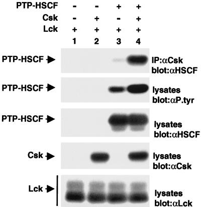
FIG. 2.
Association of Csk with PTP-HSCF in mammalian cells. Cos-1 cells were transfected (+) or not (−) with cDNAs coding for a phosphatase-inactive version of PTP-HSCF (C229S PTP-HSCF) and wild-type Csk, in the presence of a constitutively activated version of Lck (Y505F Lck). After 40 h, the association of Csk with PTP-HSCF was assessed by immunoblotting of anti-Csk immunoprecipitates (IP) with anti-PTP-HSCF (αHSCF) antibodies (first panel). Tyrosine phosphorylation of PTP-HSCF was determined by immunoblotting of total cell lysates with antiphosphotyrosine (αP.tyr) antibodies (second panel). Expression of PTP-HSCF (third panel), Csk (fourth panel), and Lck (fifth panel) was verified by immunoblotting of total cell lysates with the appropriate antibodies. The positions of PTP-HSCF, Csk, and Lck are indicated on the left. Exposures in all panels except the second panel were 2 h, and that in the second panel was 4 h.

