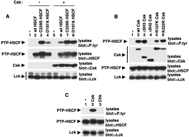
FIG. 5.
The Csk SH2 domain protects PTP-HSCF from dephosphorylation. (A) Cos-1 cells were transfected with cDNAs coding for various forms of PTP-HSCF, in the absence (lanes 1 to 4) or presence (lanes 5 to 8) of Csk. All transfections contained Y505F Lck. Tyrosine phosphorylation of PTP-HSCF was determined by immunoblotting of total cell lysates with antiphosphotyrosine (αP.tyr) antibodies (first panel). The positions of PTP-HSCF, Csk, and Lck are shown on the left. wt, wild type. Exposures in the first and second panels were 14 and 3 h, respectively, and those in the third and fourth panels were 6 h. (B) Structural requirements for induction of PTP-HSCF tyrosine phosphorylation by Csk. Cells were transfected and tested as detailed for panel A, with the exception that wild-type PTP-HSCF and various forms of Csk were utilized. The positions of PTP-HSCF, Csk, and Lck are shown on the left. Exposures in the first to fourth panels were 14, 4, 7, and 4 h, respectively. (C) Differential ability of Csk and Chk to provoke tyrosine phosphorylation of PTP-HSCF. Cells were transfected and tested as detailed for panel A, except that Csk and Chk were compared. Expression of Csk and Chk was confirmed by immunoblotting of total cell lysates with the appropriate antibodies (data not shown). The positions of PTP-HSCF and Lck are shown on the left. Exposure in the top panel was 14 h, and those in the middle and bottom panels were 6 h.

