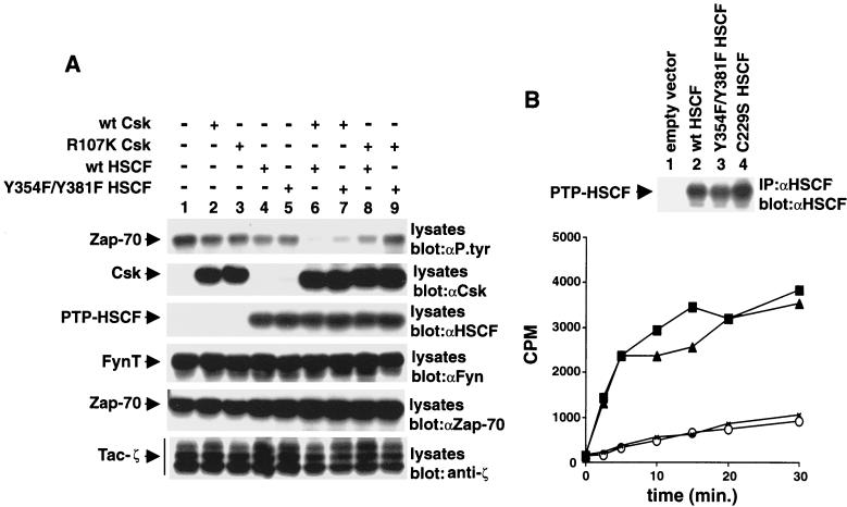
FIG. 7.
Cooperative inhibition of Src kinase-mediated signaling by Csk and PTP-HSCF. (A) Cos-1 cells were transfected with cDNAs coding for Tac-ζ, FynT, and kinase-inactive Zap-70 (K295R Zap-70), in the absence (−) or presence (+) of the indicated cDNAs. Tyrosine phosphorylation of Zap-70 was monitored by immunoblotting of total cell lysates with antiphosphotyrosine (αP. tyr) antibodies (first panel). Similar results were obtained when Zap-70 was isolated from cell lysates by immunoprecipitation (data not shown). The positions of Zap-70, Csk, PTP-HSCF, FynT, and Tac-ζ are shown on the left. wt, wild type. Exposures in the first to sixth panels were 48, 15, 5, 10, 15, and 10 h, respectively. (B) Immune complex phosphatase assays. The activities of various PTP-HSCF polypeptides were measured in an immune complex kinase assay, as outlined in Materials and Methods. The reactions were conducted for various periods of time (abscissa), and 32P-labeled myelin basic protein was used as substrate. The amount of radioactivity released in the medium is shown on the ordinate (in counts per minute [CPM]). The abundance of the PTP-HSCF proteins was verified by immunoblotting of parallel PTP-HSCF immunoprecipitates with anti-PTP-HSCF antibodies (top). Exposure, 3 h. Symbols: ○, empty vector; ×, C229S PTP-HSCF; ■, Y354F-Y381F PTP-HSCF; ▴, wild-type PTP-HSCF.

