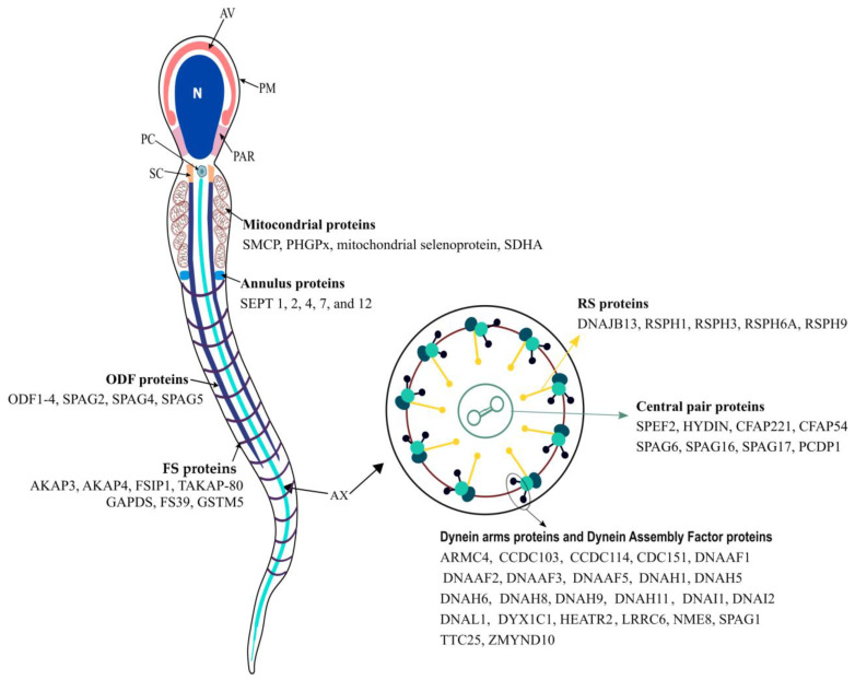
Figure 1.
Schematic view of a human sperm cell and main proteins associated with the main structures from sperm flagellum. The sperm cell is morphologically divided into two parts: the head and the flagellum. The head is composed of the plasma membrane (PM; which surrounds all sperm cells), acrosomal vesicle (AV), nucleus (N), and postacrosomal region (PAR; which connects the head to the flagellum). The sperm flagellum is further divided into four major regions, the connecting piece, the midpiece, the principal piece, and the endpiece. The connecting piece is composed mainly of the segmented columns (SCs), the proximal centriole (PC) and distal centriole, and the basal plate. For simplicity, the basal plate and distal centriole are not represented. The midpiece includes the axoneme (Ax; which extends for all flagellum), which is surrounded by the outer dense fibers (ODFs) and mitochondrial sheath (M). The Ax is composed of nine axonemal doublets microtubules (also known as the peripheral doublets) linked by the dynein regulatory complex (DRC) and connected by radial spokes (RS) to a single pair of central microtubules, which are surrounded by a fibrillar central sheath, constituting the central pair complex (CPC). The peripheral doublets are composed of microtubule A and microtubule B. From each microtubule A arise two dynein arms: the outer (ODA) and inner (IDA) dynein arms. The sperm midpiece and principal piece are separated by the annulus (An). The proximal principal piece contains the Ax surrounded by the ODF and the rings of fibrous sheath (FS). In the distal principal piece, the Ax is encircled by the FS, and the endpiece only contains the Ax, which gradually becomes disorganized and loses its 9d + 2s conformation. See text for further details.

