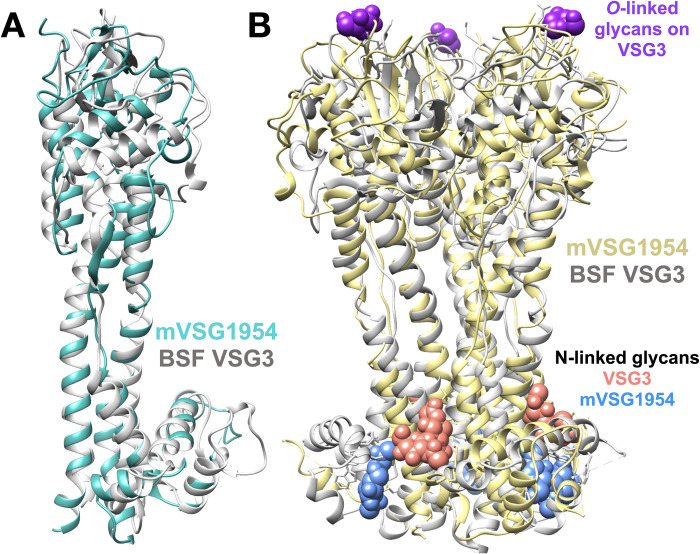
Fig 4. Crystal structure of mVSG1954.
(A) Superposition of class B mVSG1954 (aquamarine) and BSF VSG3 (grey) monomers by DeepAlign in RaptorX structure alignment server. (B) Structural superposition of the crystallographic trimers of mVSG1954 (yellow) and VSG3 (grey) performed by SSM Superpose function in COOT [37,62]. The O-linked sugar on the top lobe of VSG3 is shown in purple as a space-filling atomic representation, whereas the N-linked glycans are shown in salmon (VSG3) and blue (mVSG1954), respectively. Images of protein structures were generated and edited using CHIMERA [61].

