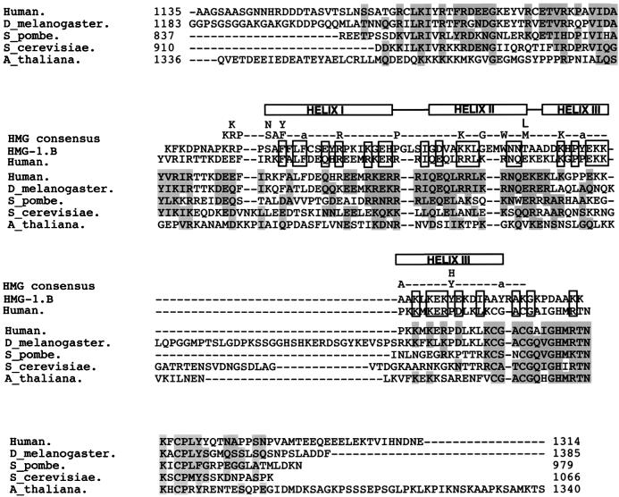
FIG. 6.
Analysis of the putative HMG domains of TAF130 homologues. Sequences corresponding to the HMG regions of several TAFs were aligned by using the program CLUSTAL W (EMBL Outstation) and compared to the original alignment between human TAF250 and the HMG-1.B (44) and to the HMG consensus (13). The three α-helices that form the HMG domain are depicted above the TAF sequences. Residues shaded in gray are conserved among the TAFs, and boxed residues are similar between human TAF250 and HMG-1.B.

