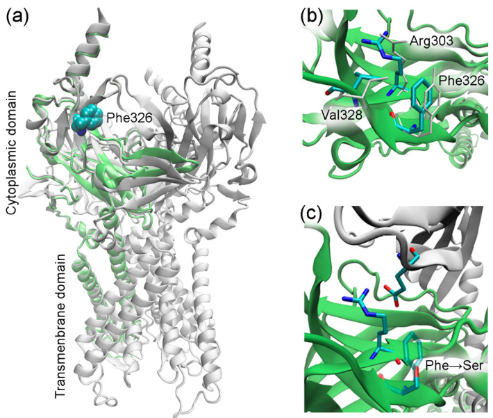
Figure 2.
Homology modeling of GIRK3. (a) Modeled GIRK3 subunit. The template GIRK2 structure (3sya, tetrameric assembly) is shown in white, and GIRK3 is shown in lime. (b) Molecular environment of the residue 326 in GIRK3 (colored by atom type) and GIRK2 (white). (c) Phe326Ser substitution in GIRK3. The GIRK2 subunit (3sya) comprising the glutamate residue is shown in white. The figure was prepared using VMD [29].

