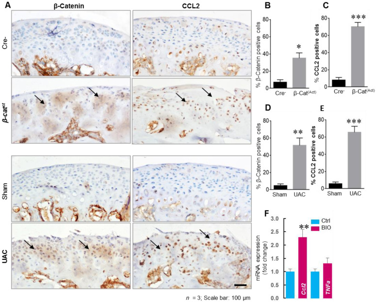
Figure 2.
β-catenin and CCL2 expression increased in TMJ condylar fibrochondrocytes in TMJ OA mice and BIO upregulated Ccl2 expression in rat condylar fibrochondrocytes. (A) TMJ tissues from β-catAct and Cre-mice were collected at the ages of 8-week-old for IHC analysis. The TMJ tissues of WT (C57BL/6) mice were also harvested 3 weeks after UAC operation for IHC analysis. Results showed that the levels of β-catenin and CCL2 increased in β-catAct mice compared to Cre-mice, and the same results were also shown in UAC-induced TMJ OA mice. Black arrows represent the typical positive expression cells in TMJ cartilage tissue. (B–E) Cells with positive staining for β-catenin and CCL2 were quantified (n = 3). Identically significant upregulation of CCL2 expression was also indicated in both β-catAct mice and UAC-induced TMJ OA mice. (F) Rat TMJ condylar fibrochondrocytes were treated with GSK-3β inhibitor, BIO, for 24 h. BIO (1 µM) significantly upregulated Ccl2 expression in TMJ condylar fibrochondrocytes and increased β-catenin protein level (Supplementary Figure S1). Statistical analysis was conducted using an unpaired t test. * p < 0.05, ** p < 0.01, *** p < 0.001.

