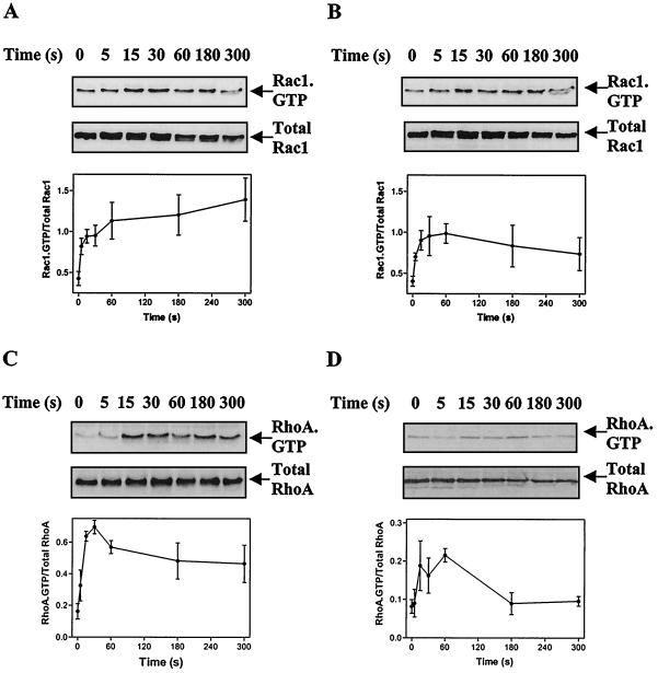
FIG. 1.
Stimulation of Rac1-GTP and RhoA-GTP in cardiac myocytes. Myocytes were exposed to 100 nM ET-1 (A and C) or 100 μM PE (B and D) for the times indicated. Rac1-GTP (A and B) or RhoA-GTP (C and D) was isolated by affinity binding assays and detected by immunoblotting (upper panels). Total Rac1 or total RhoA was also immunoblotted to ensure comparable loading (middle panels). Rac1-GTP and RhoA-GTP were quantified by scanning densitometry and were expressed relative to total Rac1 and RhoA (lower panels). Results are means ± standard errors of the means (SEM) for three (RhoA-GTP) or four (Rac1-GTP) separate myocyte preparations.

