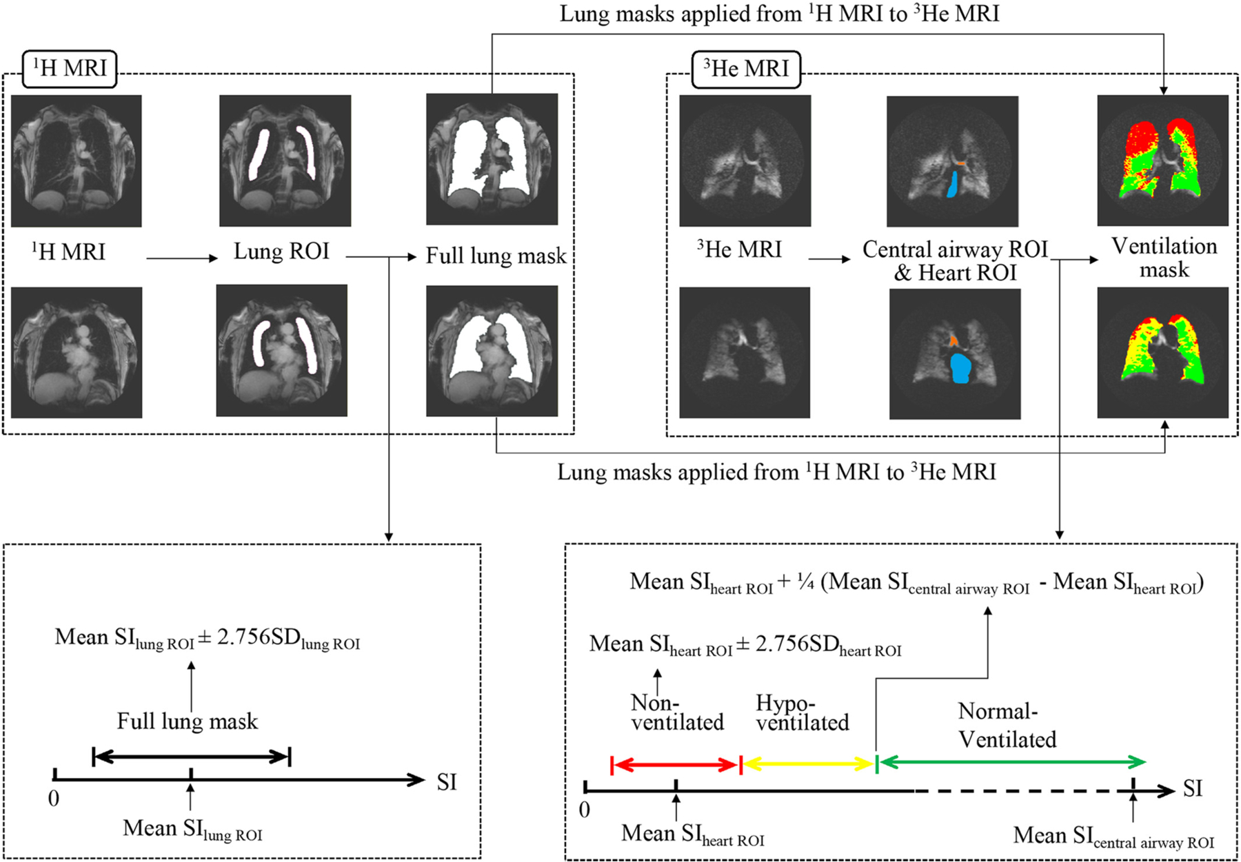
Fig. 1.

Illustration of ground truth segmentation. Full lung masks were segmented on 1H MRI and subsequently applied to 3He MRI. To determine appropriate thresholds for full lung masks and ventilation segmentations, ROIs were manually drawn inside the lungs on 1H MRI, and inside the central airways and the heart on 3He MRI. SD = standard deviation; SI = signal intensity; ROI = region of interest. Colour coding: Orange = central airway ROI; blue = heart ROI; red = non-ventilated lung regions; yellow = hypo-ventilated lung regions; green = normal-ventilated lung regions. (For interpretation of the references to colour in this figure legend, the reader is referred to the web version of this article.)
