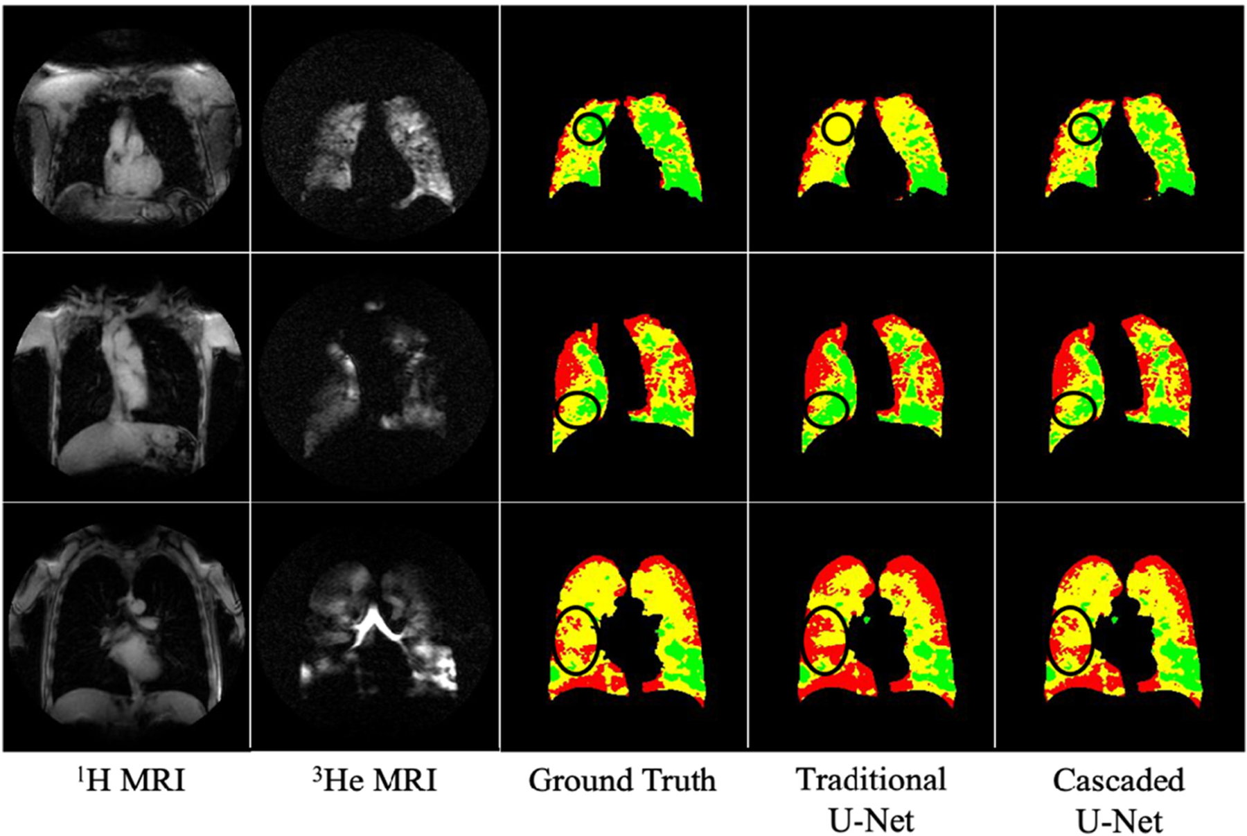
Fig. 4.

Comparison of ventilation segmentation results between traditional U-Net and cascaded U-Net. Red = non-ventilated lung region; yellow = hypo-ventilated lung region; green = normal-ventilated lung regions. The circled regions highlight higher agreement with cascaded U-Net segmentation than with traditional U-Net segmentation when compared to the ground truth. (For interpretation of the references to colour in this figure legend, the reader is referred to the web version of this article.)
