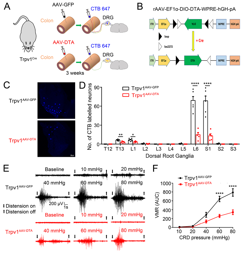
Figure 1. Virally-mediated ablation of colon-innervating TRPV1-expressing nociceptors attenuates CRD-induced VMR.

(A). Schematic representation of intracolonic injections of AAV encoding GFP or DTA into the Trpv1Cre mice followed by intracolonic injections of CTB 647 for retrograde labeling. (B). Schematic diagram illustrating the AAV-EF1α-DIO-DTA construct that expresses diphtheria toxin subunit A (DTA) in a Cre-dependent configuration to cause selective cell death in Cre-expressing neurons. (C, D). Representative images (C) and quantification (D) of retrogradely-labeled DRG neurons following injection of CTB-647 into the distal colon of TRPV1AAV-GFP and TRPV1AAV-DTA mice *P < 0.05, **p < 0.01, ****P < 0.0001, Statistical analyses by two-way ANOVA, with post hoc independent-samples t-test with FDR (False Discovery Rate) correction to compare T13, L1, L2, L5, L6, S1 and S2. (E). Representative electromyogram recordings elicited by graded CRD pressures (10, 20, 40, 60 and 80 mmHg) recorded from TRPV1AAV-GFP mice and TRPV1AAV-DTA mice. (F). Summary data showing that TRPV1AAV-DTA mice display significantly decreased VMR to CRD compared with TRPV1AAV-GFP mice at distension pressures of 60 mmHg and 80 mmHg. ****P < 0.0001, two-way ANOVA, n=5 mice per group. All data are expressed as means ± S.E.
