
FIGURE 4.
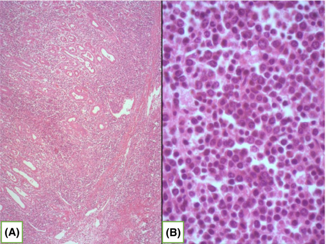
(A) Pathological examinations showing an infiltration of the gastric wall by diffuse blue proliferation HE×50; (B) Proliferation of large neoplastic lymphoid cells (usually 5 × normal lymphocytes) with amphophilic or basophilic cytoplasm, eccentric nuclei with one or more nucleoli (HE×400).