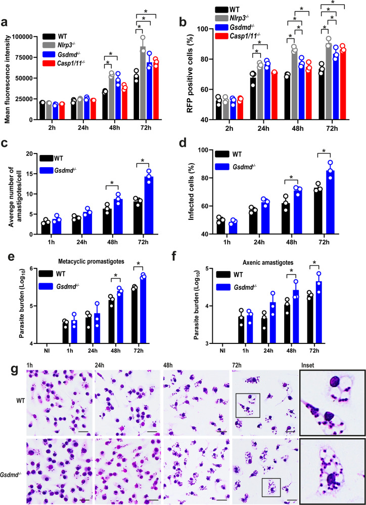
Fig. 4. GSDMD accounts for the restriction of L. amazonensis infection in bone marrow-derived macrophages (BMDMs).
a, b Flow cytometry analysis of C57BL/6 (WT), Nlrp3–/–, Gsdmd–/–, and Casp1/11–/– BMDMs infected with stationary-phase promastigotes of L. amazonensis constitutively expressing RFP. Cells were infected at an MOI 5 for 2 h, washed, and incubated for 24, 48, and 72 h. a Mean fluorescence intensity of L. amazonensis-expressing RFP. (b) The percentage of RFP-positive BMDMs. c, d Giemsa staining of C57BL/6 (WT) and Gsdmd–/– BMDMs infected with metacyclic promastigotes of L. amazonensis at an MOI 5. Cultures were infected for 1 h, washed, and incubated for 24, 48, and 72 h. c The average number of amastigotes per BMDMs. d The percentage of infected BMDMs. A total of 100 cells in each triplicate well were analyzed. e, f Parasite quantification by real-time PCR in C57BL/6 (WT) and Gsdmd–/– BMDMs infected with metacyclic promastigotes (e) and axenic amastigotes (f). g Representative images of Giemsa-stained cultures show intracellular amastigotes in BMDMs, scale bar 50 µm. Data are presented as mean values ± SD of triplicate wells. *P < 0.05 comparing the indicated groups, as determined by two-way ANOVA. Shown is one representative experiment of five independent experiments performed. Source data are provided as a Source Data file.

