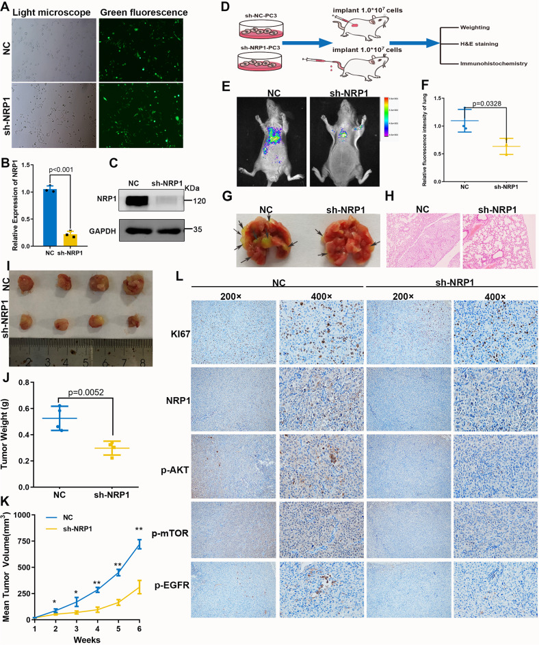
Fig. 4. NRP1 promotes PCa cell proliferation and migration in vivo.
A Green fluorescence of PC-3 stable cells. B, C qRT-PCR and immunoblot assay verify NRP1-depletion efficiency in PC-3 stable cells. D Xenograft mice models and pulmonary metastasis models were established by subcutaneously injecting LV-NC cells or LV-sh-NRP1 cells. Mice were monitored continuously for 5 weeks in xenograft mice models and 8 weeks in the pulmonary metastasis model. Then the mice were sacrificed and the tumors were dissected. E Fluorescence intensity of pulmonary metastasis is detected by imaging apparatus for small animals in vivo, F sh-NRP1 group (n = 3) significantly inhibited lung metastasis of PCa cells compared with that in the NC group (n = 3). G Lung tissue specimens show pulmonary metastatic nodules, where the arrows denote the tumors transferred to the lungs. H H&E staining of lung tissues (scale bar = 100 μm). Statistical analysis of tumor volume and weight in two groups (n = 4 in each group). I Tumor image of xenograft mice models. J NRP1 depletion significantly decreases the weight of the tumor xenograft. K NRP1 depletion significantly inhibits tumor growth. L IHC staining detects the expression of Ki67, NRP1, p-AKT, p-mTOR, and p-EGFR. Data are shown as the mean ± SD. Statistical significance was assessed using a two-tailed t test. *P < 0.01, **P < 0.001.

