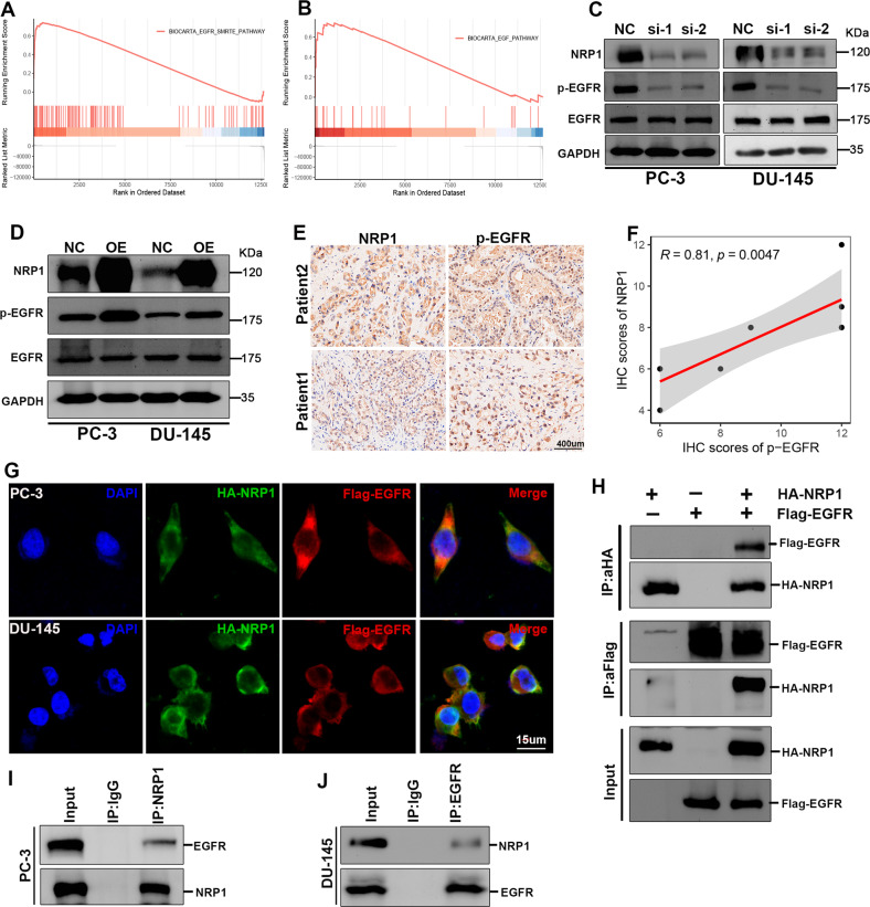
Fig. 5. NRP1 interacts with EGFR and induces EGFR phosphorylation.
A, B GSEA of TCGA-PRAD shows NRP1 is positively related to the EGFR pathway. C, D Immunoblot assay elucidates that NRP1 depletion attenuates EGFR phosphorylation level, while ectopic overexpression of NRP1 promotes EGFR phosphorylation level. E, F IHC assay and the correlation analysis represent that NRP1 protein is positively correlated with the expression of phosphorylated EGFR protein in PCa tissues (scale bar = 400 μm). G NRP1 (green) and EGFR (red) co-localization is examined by the immunofluorescence assay (scale bar = 15 μm) and the nucleus is indicated by DAPI (blue) staining. H Exogenous Co-IP assay represents NRP1 could interact with EGFR in 293T cells. I, J Endogenous Co-IP assay proves NRP1 could interact with EGFR in PC-3 and DU-145 cells. Statistical significance was assessed using a two-tailed t test.

