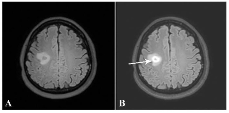
Figure 8.
Axial precontrast FLAIR (A) and postcontrast FLAIR (B) images show complete ring-enhancing lesion (arrow) with minimal surrounding vasogenic edema. No significant pressure effect is shown. A presumptive diagnosis of small tumefactive MS was made, although there were no other lesions, either in the brain or in the spinal cord. The patient was given high-dose intravenous steroid.

