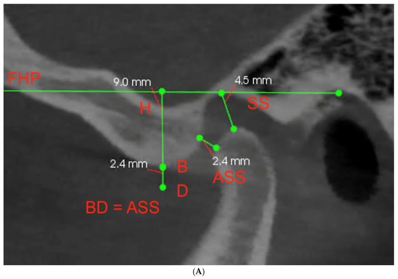
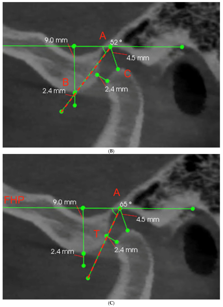
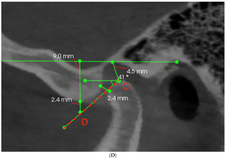
Figure 2.
(A–D) The sagittal view of the TMJ with marked landmarks and illustration of three different methods of measuring the SCGA (ASS—antero-superior space; SS—superior space; H—vertical height of the fossa; FHP—Frankfort horizontal plane; A—deepest point of the articular fossa; B—highest point of the articular eminence; T—tangent point adjacent to the articular eminence; C—highest point of the condyle; D—point below the highest point of the articular tubercle).


