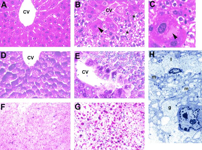
FIG. 3.
Histological analysis of the livers of H4LivKO mice. (A) Normal liver of a 45-day-old wild-type (+/+) mouse. Hepatocytes around the central vein have few vacuoles present. Shown is an H&E stain. Magnification, ×300. (B) Liver of H4LivKO mouse showing marked vacuolation of most hepatocytes (asterisks) except those around the central vein (CV). Those around the vein are eosinophilic, and one at the left side has a large intracytoplasmic pale eosinophilic inclusion (arrowhead; see panel C). Shown is an H&E stain. Magnification, ×300. (C) Centrilobular hepatocyte with large intracytoplasmic inclusion (arrowhead) in the liver of an H4LivKO mouse. Note the vacuolation of smaller hepatocytes. Shown is an H&E stain. Magnification, ×750. (D) PAS stain of normal wild-type mouse liver showing abundant PAS-positive glycogen (dark red color) in all hepatocytes around the central vein. Livers were fixed in alcoholic formalin. Magnification, ×300. (E) PAS stain of H4LivKO mouse liver showing decrease or loss of PAS-positive glycogen in most hepatocytes but abundant glycogen in centrilobular hepatocytes. Livers were fixed in alcoholic formalin. Magnification, ×300. (F) Frozen section of normal liver from a wild-type mouse, showing many small fat droplets in hepatocytes (red) throughout the liver. Shown is an oil red O stain. Magnification, ×150. (G) Frozen section of H4LivKO liver, showing abundant, large fat droplets in hepatocytes. Shown is an oil red O stain. Magnification, ×150. (H) Ultrastructure of the liver from a H4LivKO mouse showing abundant lipid (1) and mitochondria (m) in the hepatocyte at the top and areas of glycogen (g) in the hepatocyte at the bottom. Staining was with uranyl acetate and lead citrate. Magnification, ×6,000.

