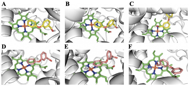
Figure 7.
PBalc and PBald binding models for the active site of P450s. The heme group is represented by green sticks, PBalc by yellow sticks and PBald by pink sticks. (A) Predicted binding model for PBalc in CYP6A36. (B) Predicted binding model for PBalc in CYP6D10. (C) Predicted binding model for PBalc in CYP4S24. (D) Predicted binding model for PBald in CYP6A36. (E) Predicted binding model for PBald in CYP6D10. (F) Predicted binding model for PBald in CYP4S24.

