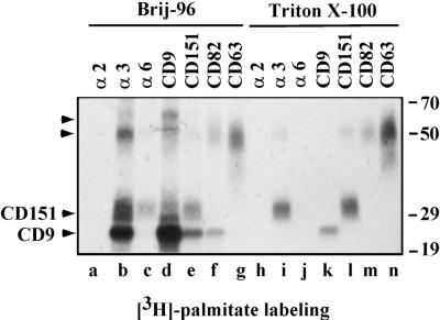
Figure 2.
Tetraspanin protein complexes. A431 cells were labeled with [3H]palmitate and then lysed in the buffers containing either 1% Brij-96 (lanes a–g) or 1% Triton X-100 (lanes h–n). Equal amounts of lysate were used for each immunoprecipitation. Antibodies for individual immunoprecipitations were A2-IIE10, A3-X8, A6-ELE, Du-All-1, 5C11, M104, and 6H1 for α2, α3, α6, CD9, CD151, CD82, and CD63, respectively. Unknown proteins of ∼60 and 50 kDa (distinct from CD63) are indicated by arrowheads. At least part of the 50-kDa protein is likely to be a CD151 dimer, as confirmed by immunoblotting for CD151 (Yang, Claas, Kraeft, Chen, Wang, Kreidberg, and Hemler, unpublished results). Note: Compared with Brij-96 extraction (lane d), Triton X-100 extraction (lane k) yielded less CD9, because of destabilization of the Du-All-1 epitope. Also, more CD151 appears in the anti-α3 lane (lane b) compared with the anti-CD151 lane (lane e), because in Brij-96 lysate, the 5C11 epitope on CD151 is likely obscured by associated proteins.

