Figure 4.
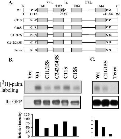
CD151 palmitoylation sites. (A) Schematic diagram of candidate CD151 palmitoylation sites. Shown are the four transmembrane domains (TM1–4), short extracellular loop (SEL), inner loop (IL), large extracellular loop (LEL), and membrane-proximal cysteine residues. Various C→S (CYS→SER) mutants are also indicated. Unless otherwise indicated, all mutants contained a GFP domain fused to the carboxy terminus. Wt, wild type. (B and C) In separate experiments, 293 cells were transiently transfected with various CD151 constructs and pulsed with [3H]palmitate (palm.). After transfection (24 h), cells were lysed in RIPA buffer and immunoprecipitated using mAb 5C11. Samples were then divided equally, resolved by SDS-PAGE, and then either dried for the detection of [3H]palmitate or transferred to nitrocellulose for blotting (Ib) with GFP polyclonal antibody.

