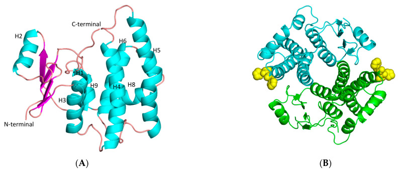
Figure 7.
(A) Representation of hGSTA1-K141H/S142H subunit. Helices and strands are shown in cyan and magenta, respectively. The helices are labeled. (B) Representation of hGSTA1-K141H/S142H dimer from the top. The two-fold axis that relates the two molecules is perpendicular to the plane of the paper. The histidine amino acids at positions 141 and 142 are shown as yellow spheres. The figures were created using PyMOL Molecular Graphics System [30].

