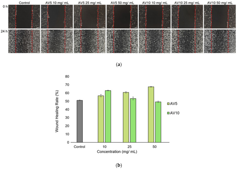
Figure 7.
Light microscope images of the L929 cell monolayer (a) after in vitro generation of a wound and treatment with different concentrations (10, 25 and 50 mg/mL) of AV5 and AV10 hydrogels, for 24 h. The cell migration in the injured area could be observed. Upper line—t = 0, lower line—after 24 h of treatment. Control was represented by untreated cells. The percentage of wound closure for L929 (b) was determined by ImageJ analysis. Data were expressed as mean values ± SD (n = 3).

