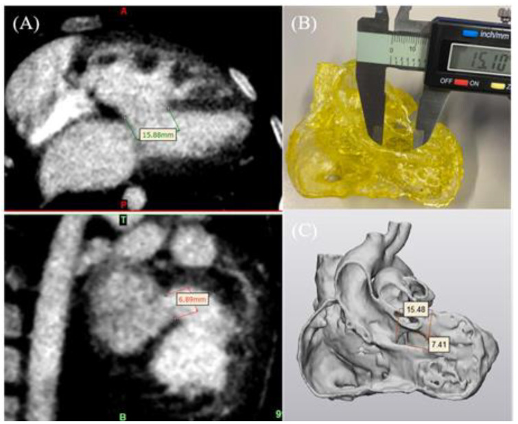
Figure 4.
3D-printed model accuracy. (A) CT imaging data in coronal and sagittal views (left top and bottom images) to measure the VSD on source imaging data. (B) Measurement of the VSD in the 3D-printed model using a digital calliper. (C) STL file measurement of the VSD in 3-Matic. VSD—ventricular septal defect. Reprinted with permission under the open access from Lee et al. [22].

