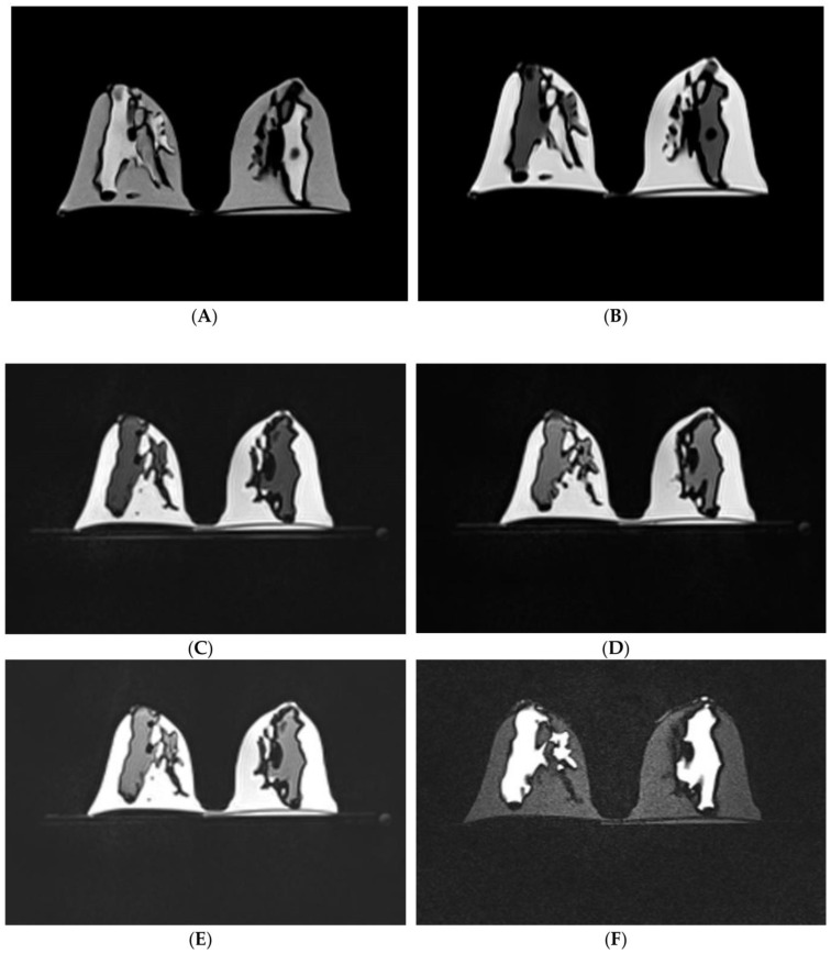
Figure 17.
MR images of 3D-printed breast model with use of six different scanning sequences. (A) Non-fat-suppressed TSE (T2WI); (B) non-fat-suppressed TSE (T1WI); (C) non-fat-suppressed TSE SPACE (T1WI); (D) fat-suppressed TSE SPACE (T1WI); (E) fat-suppressed TSE SPACE SPAIR (T1WI); (F) fat-suppressed IR/PEF-TIRM (T2WI). T1WI—T1 weighted imaging, T2WI—T2 weighted imaging, TSE—turbo (fast) spin echo, SPACE—sampling perfection with application optimized contrast using different flip angle evolution, SPAIR—spectral attenuation inversion recovery, IR—inversion recovery, PFP—partial Fourier phase, TIRM—turbo inversion recovery magnitude. Reprinted with permission under the open access from Sindi et al. [67].

