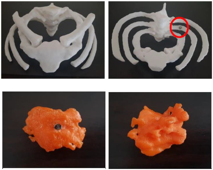
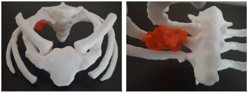
Figure 18.
3D-printed Pancoast lung cancer model and bones. (Top) row: frontal and bottom views with magnet (circled). The model was printed with polylactic acid. (Middle) row images: (Top) view of 3D-printed tumour with magnet and bottom view. (Bottom) row images: 3D-printed model with tumour and bony structure added together (frontal and superior views of the exact tumour location in the right lung apex). Reprinted with permission from Yek et al. [72].


