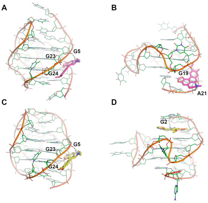
Figure 5.
Binding modes of Dicentrine to tel26 and Pu22 G-quadruplexes as predicted by molecular docking and MD simulations. (A) Docking-based binding mode of Dicentrine to the first NMR model of tel26 (PDB-ID: 2JPZ) [38]. (B) Docking-based binding mode of Dicentrine to the first NMR model of Pu22 (PDB-ID: 1XAV) [39]. (C) Representative frame extracted from the MD trajectory of tel26/Dicentrine complex. (D) Representative frame extracted from the MD trajectory of Pu22/Dicentrine complex. G-quadruplexes are shown as green cartoon and lines. Dicentrine is shown using sticks and is colored magenta in the docking poses and yellow in the poses obtained by MD simulations. Polar contacts of Dicentrine are highlighted by black dashed lines; nucleotides involved in polar interactions with Dicentrine are labeled.

