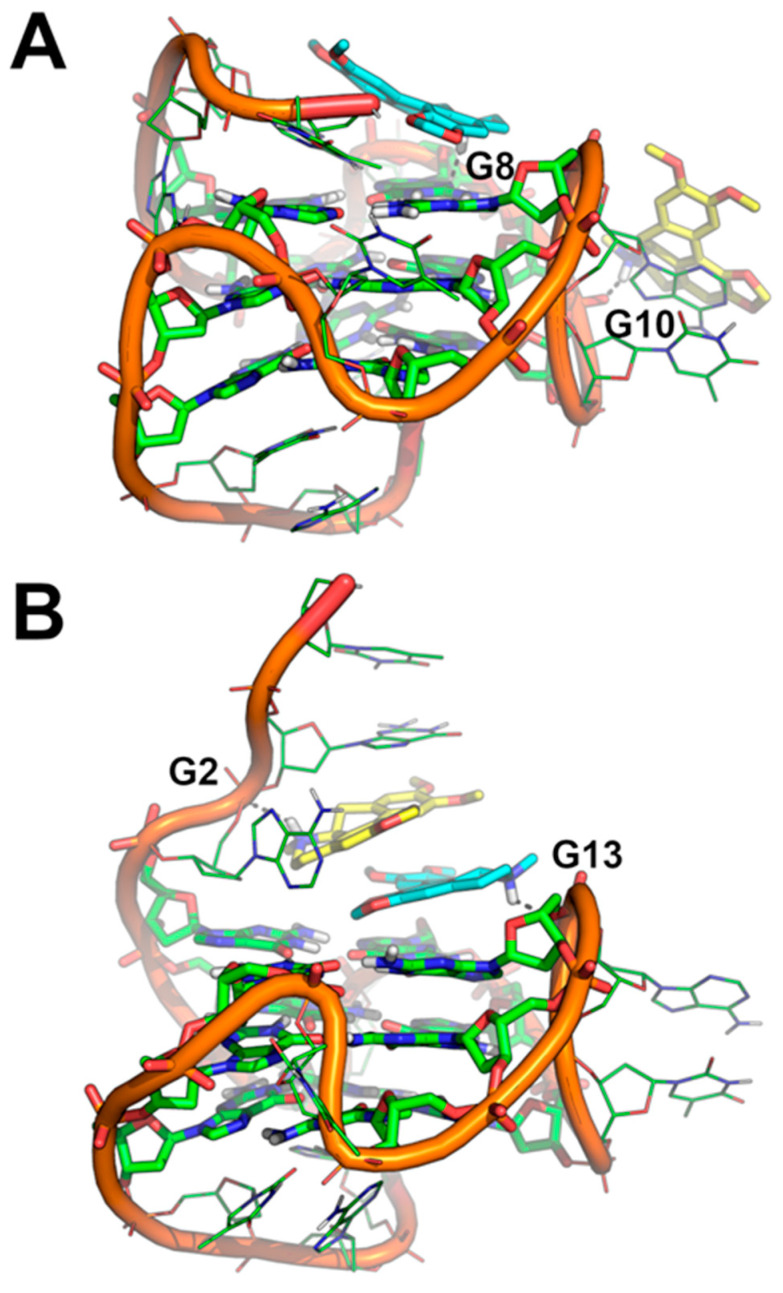
Figure 8.
(A,B) Binding mode of two molecules of Dicentrine to the Pu22 G-quadruplex investigated using two independent replicas of MD simulations. The representative frame extracted by the MD trajectories through cluster analysis is shown. Pu22 is represented as green cartoon and lines; residues forming the G-tetrads are shown as sticks. Dicentrine #1 and Dicentrine #2 are shown as sticks and colored cyan and yellow, respectively. Polar contacts are highlighted by black dashed lines; nucleotides involved in polar interactions with Dicentrine are labeled.

