Figure 12.
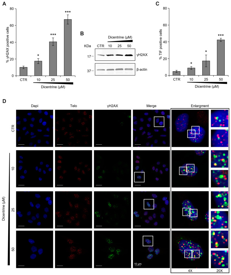
Dicentrine induces telomere-specific DNA damage. (A) HeLa cells were treated with DMSO (negative control) or with the indicated doses of Dicentrine (10, 25, 50 µM) for 24 h. Cells were processed for telomeric FISH combined with immunofluorescence, and the amount of DNA damage was evaluated. (A) Quantitative analysis of damaged cells. Results are expressed as a percentage of γH2AX positive cells over the negative control. (B) Western blot analysis of γH2AX levels. Β-actin protein levels were used as the loading control. (C) Quantitative analysis of the percentage of TIF-positive cells. Cells with at least four γH2AX-Telo colocalizations were scored as TIF-positive. (D) Representative images of confocal sections (63×) used for the quantitative analyses reported in (A,C). DAPI counterstained nuclei (blue), telomeres (red), γH2AX (green) and merged images are shown. For each experimental condition, 4× and 20× enlargements of merged fields are reported (white boxes). Scale bars (10 µm) are shown in the figures. Histograms show the mean ± SD of three independent experiments performed in triplicate. * p < 0.05; *** p < 0.001.

