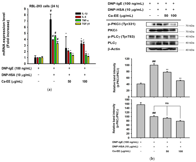
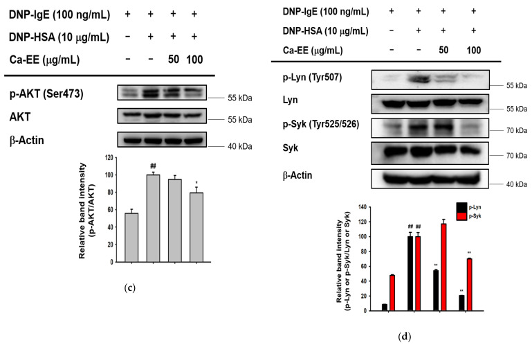
Figure 3.
Regulation by Ca-EE of inflammatory mediators in IgE- and has-treated RBL-2H3 cells. (a) Ca-EE was incubated with IgE ahasHSA for 24 h in RBL-2H3 cells. The mRNA levels were isolated from cells, and quantitative PCR was conducted. Primers for IL-1β, IL-4, TNF-α, and TGF-β1 were used. Six replicates were set in this analysis. (b–d) The effects of Ca-EE on allergic inflammatory signaling. RBL-2H3 cells were pre-incubated with Ca-EE for 30 min, and then treated with DNP-IgE (100 μg/mL) and DNP-HAS (10 μg/mL) to induce an allergic response. After 1 h, cells were harvested for immunoblotting analysis. Phosphorylated or total antibodies against PKCδ, PLCγ, AKT, Lyn, Syk and β-actin were used. β-actin was used as a loading control. The band intensity (middle and lower panels of b, and lower panels of c and d) of p-PKCδ, p-PLCγ, p-AKT, p-Lyn, and p-Syk was measured with their total forms using the Image J program, and the three replicates were used. # p < 0.05 and ## p < 0.01 compared with the normal group; * p < 0.05 and ** p < 0.01 compared with the control group. ns: not significant.


