Figure 2.
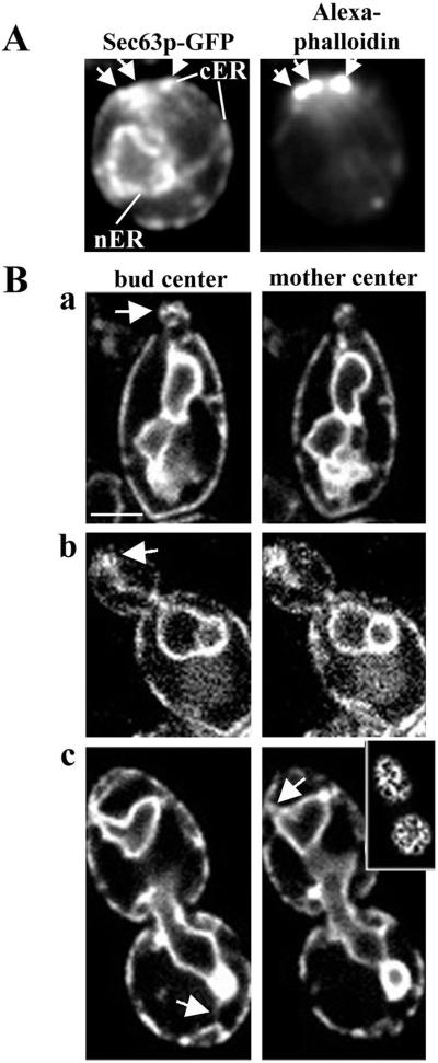
The cortical ER is enriched at the incipient bud site and in the small bud. (A) Cells expressing Sec63p-GFP were grown to midlog phase, fixed in formaldehyde, and stained with Alexa-phalloidin to visualize F-actin. Prebudded cells, with incipient bud sites marked by polarized actin patches, were imaged in a single, central focal plane. White arrows mark the position of the cortical ER (left) and the sites of actin patches at the incipient bud site (right). (B) Living cells expressing Sec63p-GFP. Cells were imaged in three dimensions with 0.3 μm between optical sections. Shown are optical sections through the center of the bud (left) and the center of the mother cell (right). Cells bearing small-sized (a), medium-sized (b), and large-sized (c) buds are shown. The inset image in c was obtained in the periphery of the cell. In a and b, white arrows mark the site of accumulation of cortical ER in the bud tip of cells in S-G2 phase. (c) In large-budded cells, after nuclear migration, the cortical ER is more evenly distributed in the bud (top); however, ER tubules (white arrowheads) make multiple contacts with the cortex, especially at the bud tip and the tip of the mother cell distal to the bud. Images were obtained with the Yokogawa confocal microscope, as described in MATERIALS AND METHODS. Bar, 1 μm.

