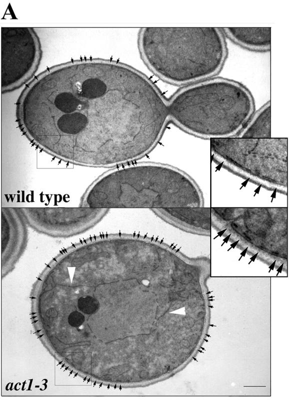
Figure 5.
Cortical ER membranes exhibit an altered pattern in an act1-3 mutant. In one EM optical section, the cortical ER appears as a linear membrane, juxtaposed to the plasma membrane. In wild-type cells (MSY106), cortical ER membranes in one thin section were approximately twice as long as those observed in act1-3 mutant cells (MSY202). Insets show twofold enlargement of boxed cortex regions. Black arrows delineate the ends, or interruptions, in the cortical ER membrane. White arrowheads show cortical ER membranes, which connect the nuclear ER and the cortical ER at the distal tip of the mother and the bud site. Bar, 1 μm.

