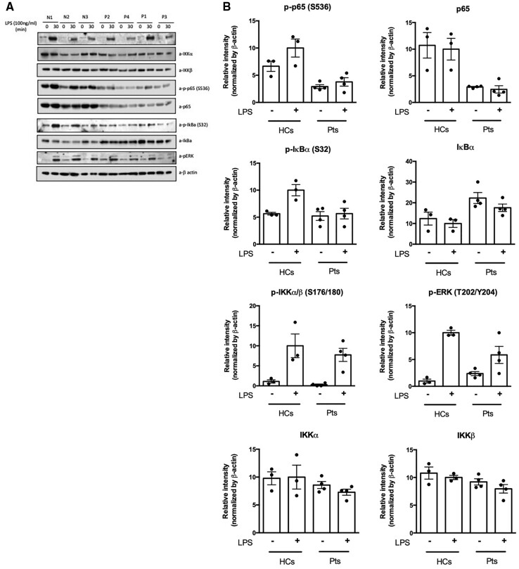
Fig. 3.
Western Blots and densitometric quantitation of immunoblotting for native and phosphorylated proteins of the canonical NFκB pathway
Total PBMCs from healthy controls and four patients were stimulated with LPS (100 ng/mL) for 30 min. (A) The Western Blot images were acquired with a C-Digit scanner, and data were analysed using Image Studio software (Li-Cor). (B) The proteins of the NFkB canonical pathway from the Western Blot were quantified using densitometry, and the levels of proteins were normalized to β-actin. The relative phosphorylation and protein levels are shown. The values are expressed as the mean (s.d.).

