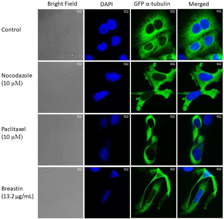
Figure 7.
Enhanced microtubule network in Breastin-treated U2OS cells. Micrographs of cells fixed with 4% paraformaldehyde were taken 24 h post-treatment with DMSO, Breastin (13.2 µg/mL), paclitaxel (10 µM), and nocodazole (10 µM). DAPI was used to stain nuclei. Images were taken at 40× magnification (scale bars = 10 µm) with the AF7000 widefield fluorescence microscope.

