
Figure 6.
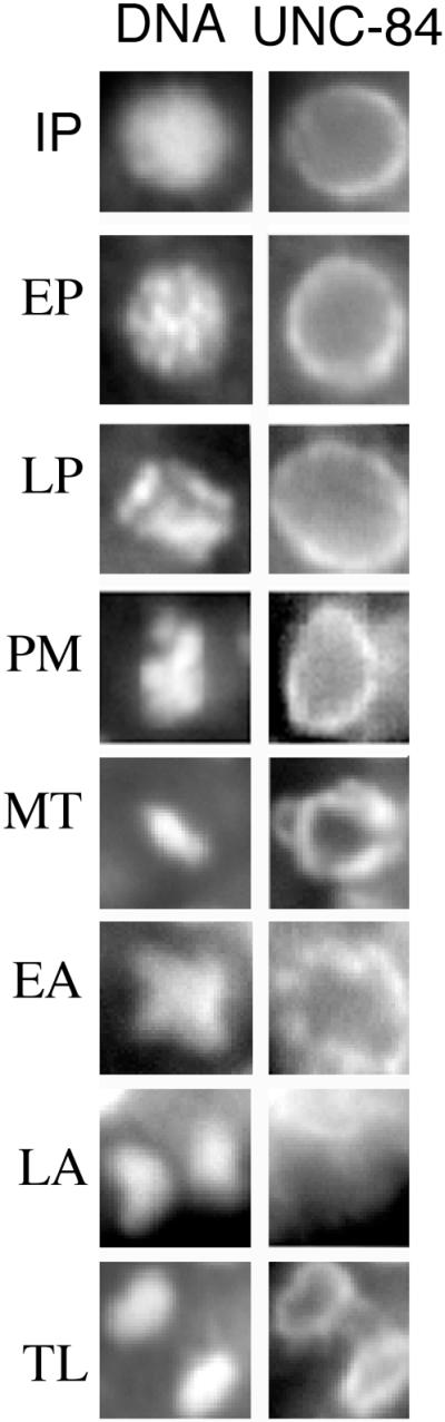
UNC-84 localization during mitosis. Wild-type embryos (100–300 cells) were stained with rat anti–UNC-84 antibodies (right panels) and Hoechst 33258 to stain DNA (left panels), viewed by fluorescence microscopy and photographed at different stages of the cell cycle. A representative nucleus from each stage is shown: IP, interphase; EP, early prophase; LP, late prophase; PM, prometaphase; MT, metaphase; EA, early anaphase; LA, late anaphase and TL, telophase. Nuclear staining was detected in all stages of mitosis except late anaphase, consistent with other nuclear membrane markers (Lee et al., 2000).