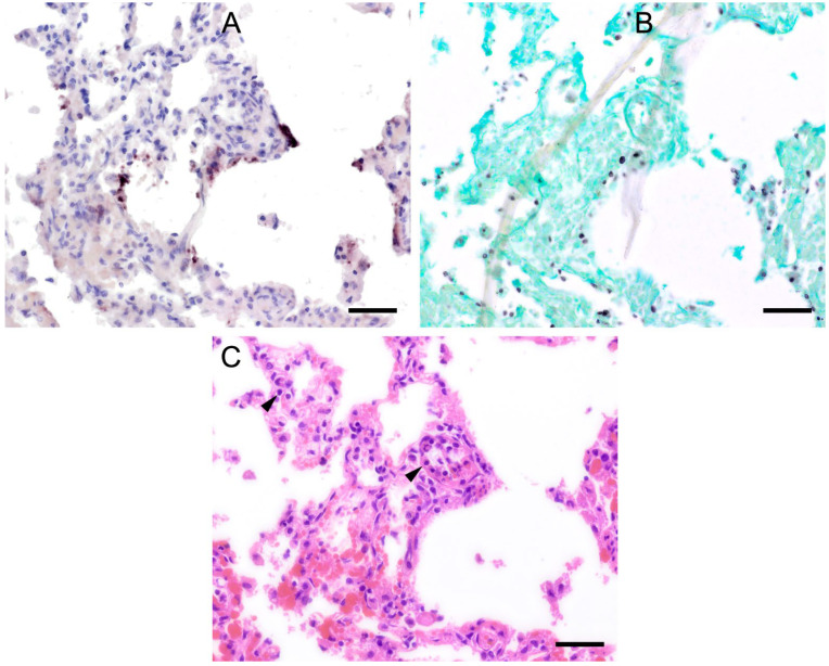
Figure 5.
Adjacent sections from the same lung area of a wild boar with moderate colonization with Pneumocystis spp. are shown in comparison after ISH, GMS, and H&E staining. (A) ISH labels Pneumocystis organisms attached to the alveolar walls of the entire lung tissue section. Bar = 40 μm; (B) GMS labels exclusively cystic forms. Bar = 40 μm; (C) H&E staining reveals an interstitial pneumonia with moderate lymphocytic infiltration (arrowheads). Bar = 40 μm.

