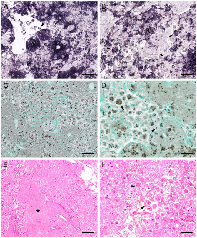
Figure 10.
Sections from the lung of a dog with severe PCP. (A,B) Large areas with numerous developmental stages are labeled by ISH. Signals are present free (asterisks) and within cells with the morphology of alveolar macrophages (arrows). (A) Bar = 80 μm; (B) Bar = 40 μm; (C) GMS labels only cystic stages black. Trophozoites largely remain unstained. Bar = 80 μm; (D) Asci are present free (arrowhead) and phagocytized within cells with the morphology of alveolar macrophages (arrow). Bar = 40 μm; (E) Severely distended alveolar spaces are diffusely filled with myriads of eosinophilic, foamy, or spherical structures (asterisk). Bar = 80 μm; (F) Variably sized cells with the morphology of macrophages containing Pneumocystis organisms (arrows). Bar = 40 μm.

