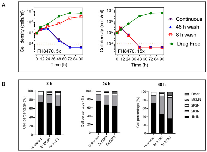
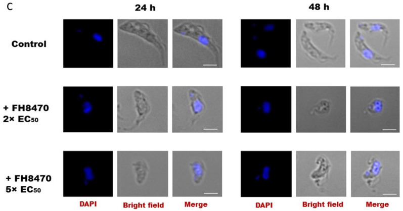
Figure 11.
The effect of tubercidin analog FH8470 on growth, cell cycle and morphology of T. congolense. (A) Manual cell count of cultures grown in the presence or absence of 5 × or 15 × EC50 of tubercidin analog FH8470. For short exposure, cultures were washed through centrifugation and resuspended in fresh medium. The dotted brown line indicates the detection limit, being 104 cells/mL. For the purpose of this graphical representation, where no cells were observed in the counting chamber, the value of 5 × 10³ or 4 × 10³ was entered. (B) Effect of nucleoside analog FH8470 on the cell cycle progression in T. congolense. Cells were seeded at 105 cells/mL in growth medium with or without test drug at the desired concentration. At each predetermined period, approximately 106 cells were harvested, transferred to a glass slide and fixed. The slide was then DAPI-treated and covered with a coverslip, and images were acquired using Olympus IX71 DeltaVision Core System fluorescent microscope (Applied Precision, GE, Rača, Slovakia). The configurations of nucleus and kinetoplast for at least 300 cells were observed in each group; 1K1N = one kinetoplast and one nucleus; 2K1N = two kinetoplasts and two nuclei; 2K2N = two kinetoplasts and two nuclei; MKMN = multiple kinetoplasts and/or nuclei; other = other aberrations such as no nucleus or no kinetoplast. Graphs show percentage mean ± SEM of three independent determinations. (C) Images of T. congolense cells exposed to nucleoside analog FH8470 for 24 h or 48 h. Test drug at desired concentration was added to BSF T. congolense culture followed by incubation for 24 h or 48 h. Cells were harvested at predetermined point, applied to a glass slide, fixed and treated with DAPI before the slide was covered with a coverslip. Images were acquired using a Delta Vision Core fluorescent microscope and analyzed using ImageJ software. Treated culture showed higher percentage of cells with conjoined nuclei and two kinetoplasts, often accompanied by mottled cell surface and loss of slender BSF shape. White bar represents 3 µM.


