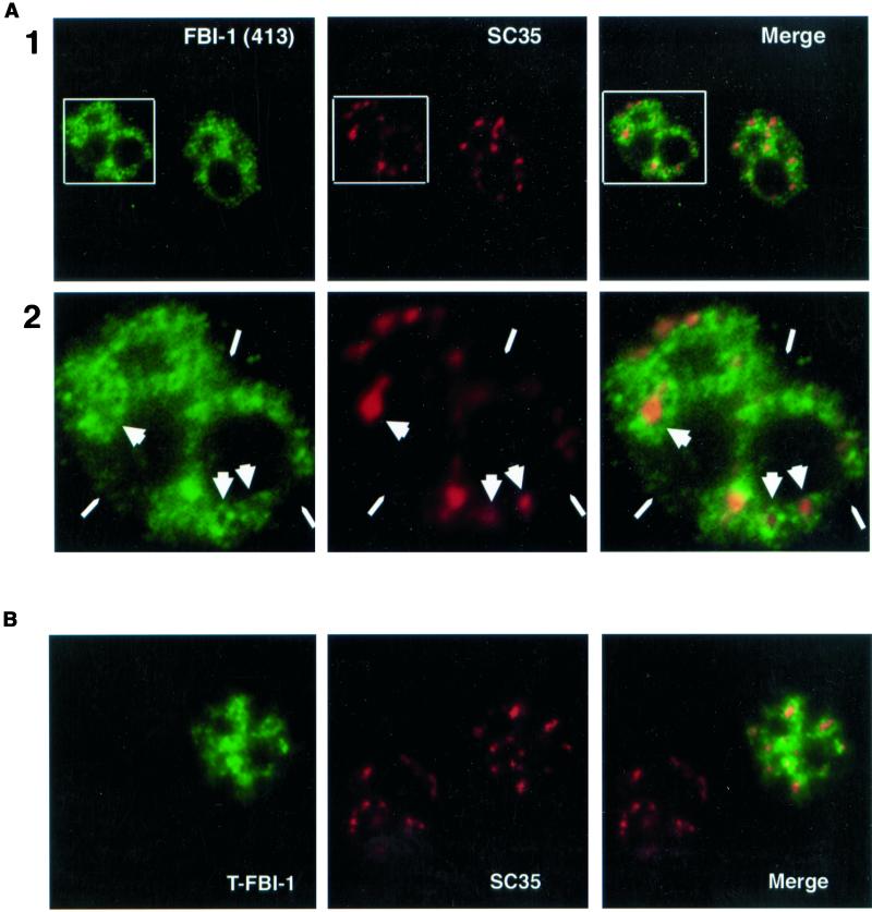
Figure 5.
Less-soluble FBI-1 displays peripheral-speckle localization. Epifluorescent image of prefixation CSK extracted HeLa cells. Less-soluble endogenous FBI-1 (green) shows strong signal surrounding and connecting the nuclear speckles (red) but shows a much weaker signal at the center of many speckles (large arrowheads) and from regions of the nucleus that have no speckles (narrow arrowheads). FBI-1 was detected with 413 primary antibody or with another rabbit polyclonal antibody specific for FBI-1 (415, our unpublished results) and FITC-GAR secondary antibody. Nuclear speckles were detected with SC35 primary and TR-GAM secondary antibodies. (B) Confocal image of prefixation CSK extracted Hela cells transfected with 2 μg of T7-FBI-1 (FL). T7-FBI-1 (green) signal is enhanced at the periphery of the SC35 nuclear speckles (red). T7-FBI-1 was detected with a mouse monoclonal primary antibody (T7 epitope antibody, Novagen) and FITC-GAM secondary antibody. Nuclear speckles were detected as in A.

