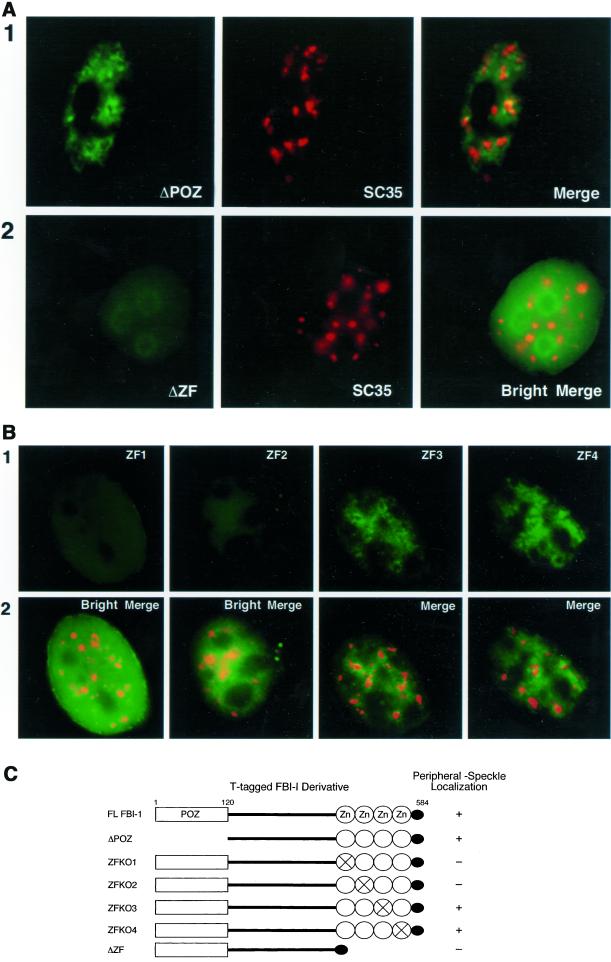
Figure 6.
The DNA-binding zinc fingers 1 and 2 are required for less-soluble FBI-1's peripheral-speckle pattern of localization. Epifluorescent image of prefixation extracted HeLa cells transfected with 2 μg of the T-FBI-1 mutant-expressing plasmids ΔPOZ (A, panel 1), ΔZF (A, panel 2), ZF1 (B, column 1), ZF2 (B, column 2), ZF3 (B, column 3), and ZF4 (B, column 4). (A) Less-soluble T-FBI-1 ΔPOZ (panel 1, green) shows a peripheral-speckle pattern of localization (Merge), whereas T-FBI-1 ΔZF (panel 2, green) shows an aberrant homogeneous pattern of localization with an increased signal from the nucleoli and no increase in signal near the nuclear speckles (box 2, red and box 3). The T-tagged FBI-1 mutants were detected as T-FBI-1 in Figure 4. The nuclear speckles were detected as in Figure 5. Bright images result from 8× exposure times. (B) Less-soluble T-FBI-1 ZF3 and ZF4 (columns 3 and 4, respectively, green) also show a peripheral-speckle pattern of localization; however, T-FBI-1 ZF1 and ZF2 show aberrant homogeneous patterns of localization (columns 1 and 2, respectively, green). The T-tagged point mutants were detected as T-FBI-1 in Figure 6. Bright images result from 10× exposure times. (C) Summary of results from A and B.

