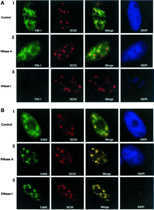
Figure 7.
Less soluble FBI-1's peripheral-speckle pattern is dependent on cellular DNA but not on cellular RNA. Epifluorescent image of prefixation extracted HeLa cells treated for 30 min at 37°C with either RNase A (A and B, panel 2), DNase I (A and B, panel 3), or buffer (A and B, panel 1). (A) FBI-1's (green) peripheral-speckle pattern is little disturbed by treatment with buffer (panel 1) or RNase A (panel 2); however, less-soluble FBI-1 signal is drastically reduced upon DNase I treatment (panel 3, column 1), whereas the nuclear speckles (red) are undisturbed. Cdk9, and nuclear speckles were detected as in Figure 5. FBI-1 was detected as in Figure 4. (B) Less soluble Cdk9's (green) localization pattern is relatively undisturbed by all three treatments (column 1) and still colocalizes (yellow) with nuclear speckles (red). DNase I treatment results in a strong reduction of DNA content in the cell as judged by DAPI staining (A and B, blue)

