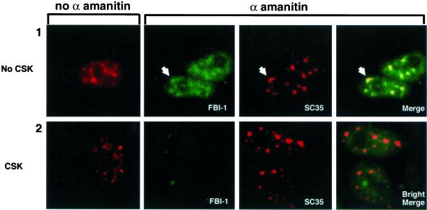
Figure 8.
Total FBI-1 is redistributed to enlarged and rounded nuclear speckles, but the less-soluble FBI-1 population and peripheral-speckle pattern is drastically reduced upon inhibition of transcription. Epifluorescent images of nonextracted (panel 1) and prefixation extracted (panel 2) HeLa cells either untreated (column 1) or treated for 5 h with 50 μg/ml of α-amanitin (columns 2–4). Total FBI-1 (panel 1, green) is redistributed into enlarged nuclear speckles (red and yellow). Note that unlike in non–α-amanitin–treated cells (Figure 4C), the FBI-1 signal foci match the shape, size, and relative intensity of the nuclear speckles even in irregularly shaped foci (arrowhead). However, less-soluble FBI-1 signal is almost completely lost (panel 2, column 2). A six times longer exposure (panel 2, column 4, Bright) shows that the little FBI-1 that is left after extraction does not show a peripheral-speckle pattern of localization. Nuclear speckles were detected as in Figure 5. FBI-1 was detected as in Figure 4.

