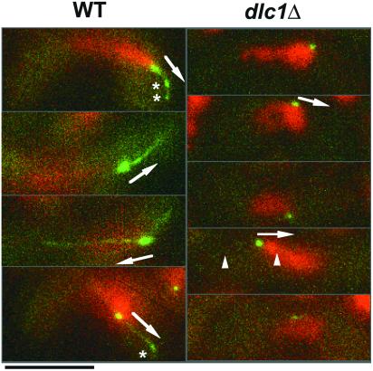
Figure 7.
Reduction in localized GFP-Dhc1 in meiotic prophase nuclei in the dlc1Δ mutant. Strains MB125 (WT) and F73-3A (dlc1Δ) were observed without fixation. Representative images obtained from different cells are shown. Green, GFP-Dhc1; red, DNA. Arrows indicate the direction of nuclear movement. Asterisks indicate presumed microtubule anchors on the cell cortex; arrowheads indicate GFP signals along cytoplasmic microtubules in the dlc1 mutant. Bar, 5 μm.

