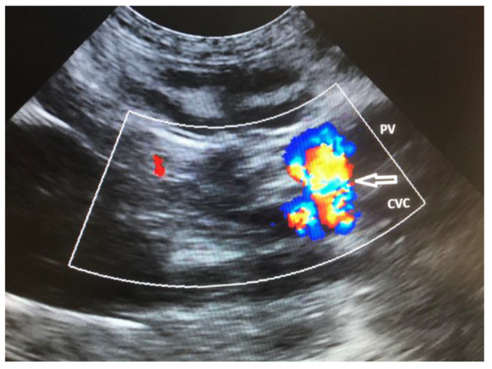
Figure 4.
Sagittal plane ultrasound color flow Doppler image of the cranial abdomen in a dog showing the presence of an extrahepatic communication (arrow) of the portal vein (PV) with the caudal vena cava (CVC). There is turbulent blood flow within the shunt vessel indicated by the mosaic pattern of colors.

