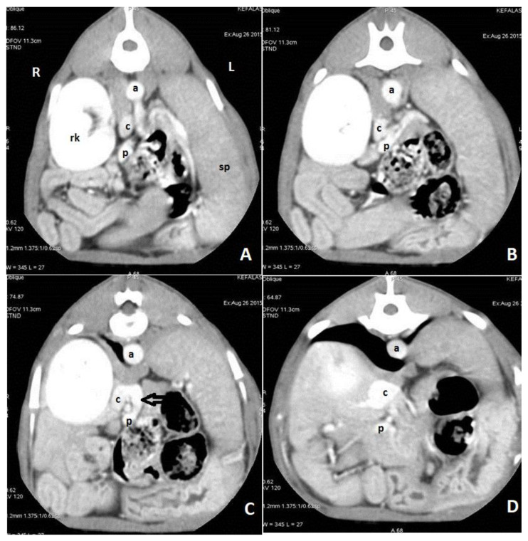
Figure 7.
Axial portal phase angiographic images of a dog with an extrahepatic portocaval shunt. Images are ordered from caudal to cranial (A–D). A shunt vessel (black arrow) arising from the portal vein (p) enters into the caudal vena cava (c). There is dilation of the caudal vena cava after the insertion of the shunt vessel. (a: aorta; rk: right kidney; sp: spleen: R: right side; L: left side).

