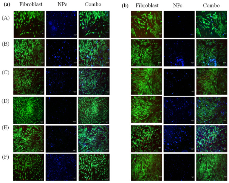
Figure 5.
In vitro cell uptake of fluorescent nanoparticles of day 1 (a) and day 3 (b) with 4-AP (10 µg/mL) and IND (50 µg/mL) in drug media solution (n = 5). The left columns of each panel (A,B) show live/dead staining images of fibroblast cells. The middle column shows fluorescent nanoparticles (A) CA-PBC, (B) CA-PBC-IND, (C) CA-PBC-4-AP, (D) PCL-PEG-PBC, (E) PCL-PEG-PBC-IND, and (F) PCL-PEG-PBC-4-AP. The right columns for each panel (a,b) show combined (Combo) images. The layover images show the presence of nanoparticles taken by the cells as well as in the culture media. The fibroblast cells maintained morphology with no evident cell death by the nanoparticle addition from Day 1. Day 3 shows some cell death for CA-PBC-IND compared to CA-PBC-4-AP (scale bar 100 µm).

