Figure 3.
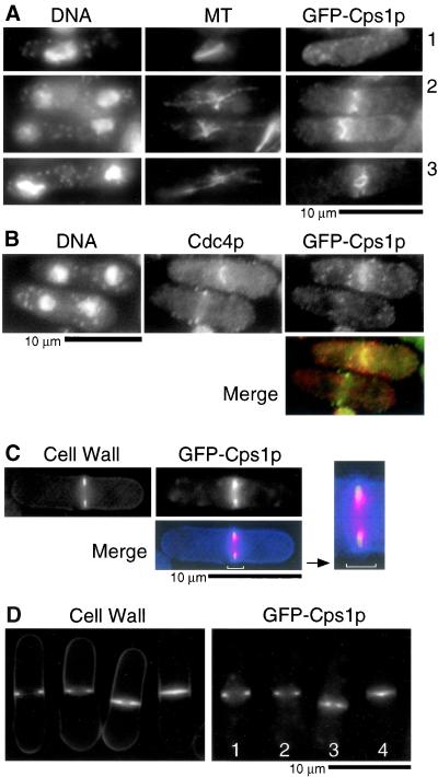
GFP-Cps1p localizes to the medial actomyosin ring late in anaphase. (A) Cells bearing GFP-cps1+ as the chromosomal copy were stained with DAPI, TAT1, and anti-GFP antibodies to visualize DNA, microtubules (MT), and GFP-Cps1p. (B) Cells were stained for DNA, Cdc4p, and GFP-Cps1p with DAPI, rabbit anti-Cdc4p antibodies, and mouse anti-GFP antibodies, respectively. In the merged image Cps1p is in red and Cdc4p in green. (C) GFP-Cps1p–expressing cells were stained for 1,3-β-glucan with aniline blue. In the merged image Cps1p is in red and septum in blue. (D) The localization of 1,3-β-glucan (aniline blue staining) and GFP-Cps1p is shown in cells at different stages of cytokinesis. Cells marked 1–4 represent those with GFP-Cps1p at different stages of constriction, with 1 being least advanced and 4 being most advanced.

