Figure 6.
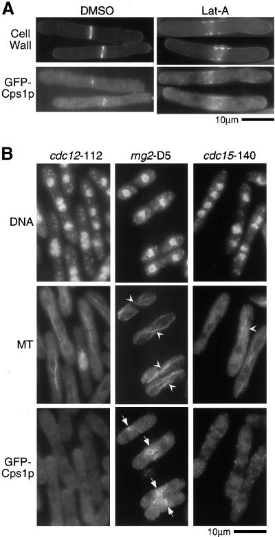
The actomyosin ring serves as a spatial landmark for Cps1p assembly at the division site. (A) cdc25-22 cells expressing GFP-Cps1p were synchronized at 36°C, released to 24°C in medium containing either 100 μM latrunculin A (LatA) or DMSO (solvent control), and stained with aniline blue. (B) Cells bearing cdc12-112, rng2-D5, or cdc15-140 were grown at nonpermissive temperature (36°C) and stained for DNA, microtubules (MT), and GFP-Cps1p with DAPI, TAT1 antibodies, and anti-GFP antibodies, respectively. Arrowheads and arrows indicate cells with microtubule configuration resembling postanaphase array and GFP-Cps1p medial structures, respectively.

