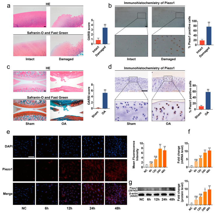
Figure 1.
Piezo1 was up-regulated in the articular cartilage of OA patients and rats. (a,c) Representative images of HE, Safranin-O/Fast Green staining of intact and damaged areas in OA patients’ articular, Sham, and OA rat cartilage, and OARSI scores (n = 5). (b,d) Immunohistochemistry staining of intact and damaged areas in OA patients’ articular, Sham, and OA rat cartilage, and the percentage of Piezo1 positive cells (n = 5). (e) Immunofluorescence staining and quantified results of Piezo1 in chondrocytes exposed to mechanical strain for 0 h, 6 h, 12 h, 24 h, and 48 h (n = 3). (f,g) RT-qPCR and Western blot analysis of Piezo1 in chondrocytes exposed to mechanical strain for 0 h, 6 h, 12 h, 24 h, and 48 h. * p < 0.05; ** p < 0.01; ns, no significant differences. Mann–Whitney U test for (a,c), Student’s t-test for (b,d), one-way ANOVA with Bonferroni’s test for (e–g); scale bar: 100 μm.

